
Journal of Frontiers of Computer Science and Technology ›› 2022, Vol. 16 ›› Issue (3): 683-691.DOI: 10.3778/j.issn.1673-9418.2010061
• Graphics and Image • Previous Articles Next Articles
GU Penghui, XIAO Zhiyong+( )
)
Received:2020-10-23
Revised:2021-01-05
Online:2022-03-01
Published:2021-01-28
About author:GU Penghui, born in 1997, M.S. candidate, student member of CCF. His research interest is medical image processing.Supported by:
谷鹏辉, 肖志勇+()
通讯作者:
+ E-mail: zhiyong.xiao@jiangnan.edu.cn作者简介:谷鹏辉(1997—),男,河南许昌人,硕士研究生,CCF学生会员,主要研究方向为医学图像处理。基金资助:CLC Number:
GU Penghui, XIAO Zhiyong. Application of Improved U-Net in Retinal Vessel Segmentation[J]. Journal of Frontiers of Computer Science and Technology, 2022, 16(3): 683-691.
谷鹏辉, 肖志勇. 改进的U-Net在视网膜血管分割上的应用[J]. 计算机科学与探索, 2022, 16(3): 683-691.
Add to citation manager EndNote|Ris|BibTeX
URL: http://fcst.ceaj.org/EN/10.3778/j.issn.1673-9418.2010061

Fig.1 Five layers of DenseNet modules
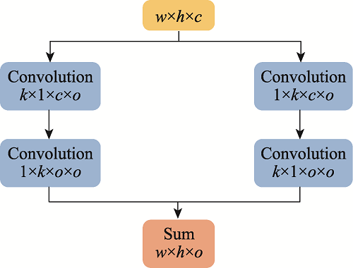
Fig.2 Global convolutional network

Fig.3 Boundary refinement
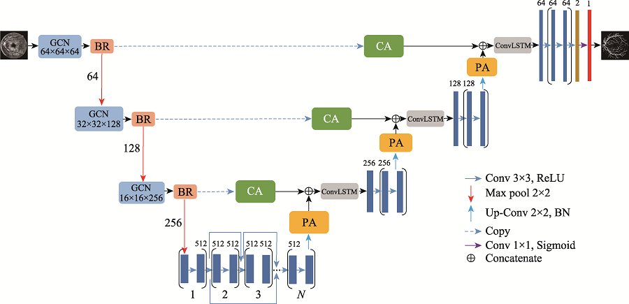
Fig.4 Network mechanism

Fig.5 Position attention module
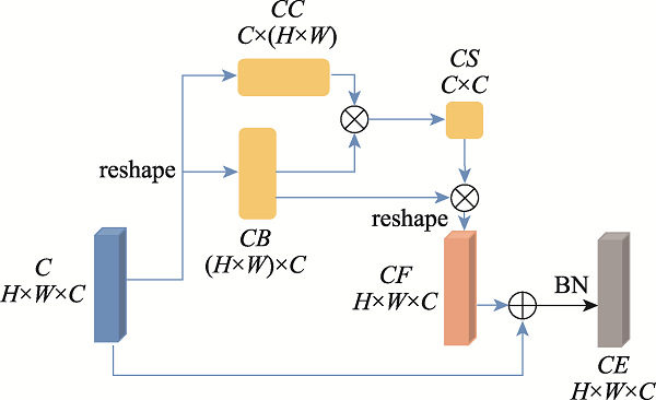
Fig.6 Channel attention module

Fig.7 Preprocessing results
| Dataset | Method | Se | Ac | AUC | F1-Score |
|---|---|---|---|---|---|
| DRIVE | 未预处理 | 81.78 | 96.99 | 98.74 | 82.67 |
| 预处理 | 83.24 | 96.99 | 98.77 | 82.91 | |
| 未后处理 | 79.74 | 97.06 | 98.77 | 82.64 | |
| 后处理 | 83.24 | 96.99 | 98.77 | 82.91 | |
| CHASE_DB1 | 未预处理 | 80.12 | 97.48 | 99.00 | 83.48 |
| 预处理 | 81.49 | 97.51 | 99.01 | 83.55 | |
| 未后处理 | 79.56 | 97.54 | 99.01 | 83.19 | |
| 后处理 | 81.49 | 97.51 | 99.01 | 83.55 |
Table 1 Comparison of pre/post processing results %
| Dataset | Method | Se | Ac | AUC | F1-Score |
|---|---|---|---|---|---|
| DRIVE | 未预处理 | 81.78 | 96.99 | 98.74 | 82.67 |
| 预处理 | 83.24 | 96.99 | 98.77 | 82.91 | |
| 未后处理 | 79.74 | 97.06 | 98.77 | 82.64 | |
| 后处理 | 83.24 | 96.99 | 98.77 | 82.91 | |
| CHASE_DB1 | 未预处理 | 80.12 | 97.48 | 99.00 | 83.48 |
| 预处理 | 81.49 | 97.51 | 99.01 | 83.55 | |
| 未后处理 | 79.56 | 97.54 | 99.01 | 83.19 | |
| 后处理 | 81.49 | 97.51 | 99.01 | 83.55 |

Fig.8 Post-processing results
| Method | DRIVE | CHASE_DB1 | ||||||||
|---|---|---|---|---|---|---|---|---|---|---|
| Se | Ac | AUC | F1-Score | Se | Ac | AUC | F1-Score | |||
| U-Net[ | 75.37 | 95.31 | 97.55 | 81.42 | 82.88 | 95.78 | 97.72 | 77.83 | ||
| GCN+BR_U-Net | 82.81 | 96.98 | 98.73 | 82.77 | 81.25 | 97.49 | 98.99 | 83.50 | ||
| GCN+BR_ConvLSTM_U-Net | 83.10 | 96.98 | 98.75 | 82.84 | 81.37 | 97.50 | 98.97 | 83.52 | ||
| GCN+BR_ConvLSTM_CA+PA_U-Net | 83.24 | 96.99 | 98.77 | 82.91 | 81.49 | 97.51 | 99.01 | 83.55 | ||
Table 2 Comparison of segmentation algorithms of several improved strategies %
| Method | DRIVE | CHASE_DB1 | ||||||||
|---|---|---|---|---|---|---|---|---|---|---|
| Se | Ac | AUC | F1-Score | Se | Ac | AUC | F1-Score | |||
| U-Net[ | 75.37 | 95.31 | 97.55 | 81.42 | 82.88 | 95.78 | 97.72 | 77.83 | ||
| GCN+BR_U-Net | 82.81 | 96.98 | 98.73 | 82.77 | 81.25 | 97.49 | 98.99 | 83.50 | ||
| GCN+BR_ConvLSTM_U-Net | 83.10 | 96.98 | 98.75 | 82.84 | 81.37 | 97.50 | 98.97 | 83.52 | ||
| GCN+BR_ConvLSTM_CA+PA_U-Net | 83.24 | 96.99 | 98.77 | 82.91 | 81.49 | 97.51 | 99.01 | 83.55 | ||
| Method | Year | Se/% | Ac/% | AUC/% | F1-Score/% |
|---|---|---|---|---|---|
| R2U-Net[ | 2018 | 77.92 | 95.56 | 97.84 | 81.71 |
| U-Net[ | 2018 | 75.37 | 95.31 | 97.55 | 81.42 |
| LadderNet[ | 2018 | 78.56 | 95.61 | 97.93 | 82.02 |
| DEU-Net[ | 2019 | 79.40 | 95.67 | 97.72 | 82.70 |
| AG-Net[ | 2019 | 81.00 | 96.92 | 98.56 | N.A |
| 吴鑫鑫等人[ | 2019 | 81.92 | 96.95 | 97.82 | N.A |
| 吕晓文等人[ | 2020 | 80.62 | 95.47 | 97.39 | N.A |
| AttR2U-Net[ | 2020 | 80.28 | 96.89 | 98.41 | N.A |
| Zhang等人[ | 2020 | 81.51 | 96.95 | 98.63 | N.A |
| RVSeg-Net[ | 2020 | 81.07 | 96.81 | 98.17 | N.A |
| Proposed | 2020 | 83.24 | 96.99 | 98.77 | 82.91 |
Table 3 Results of different algorithms on DRIVE dataset
| Method | Year | Se/% | Ac/% | AUC/% | F1-Score/% |
|---|---|---|---|---|---|
| R2U-Net[ | 2018 | 77.92 | 95.56 | 97.84 | 81.71 |
| U-Net[ | 2018 | 75.37 | 95.31 | 97.55 | 81.42 |
| LadderNet[ | 2018 | 78.56 | 95.61 | 97.93 | 82.02 |
| DEU-Net[ | 2019 | 79.40 | 95.67 | 97.72 | 82.70 |
| AG-Net[ | 2019 | 81.00 | 96.92 | 98.56 | N.A |
| 吴鑫鑫等人[ | 2019 | 81.92 | 96.95 | 97.82 | N.A |
| 吕晓文等人[ | 2020 | 80.62 | 95.47 | 97.39 | N.A |
| AttR2U-Net[ | 2020 | 80.28 | 96.89 | 98.41 | N.A |
| Zhang等人[ | 2020 | 81.51 | 96.95 | 98.63 | N.A |
| RVSeg-Net[ | 2020 | 81.07 | 96.81 | 98.17 | N.A |
| Proposed | 2020 | 83.24 | 96.99 | 98.77 | 82.91 |
| Method | Year | Se/% | Ac/% | AUC/% | F1-Score/% |
|---|---|---|---|---|---|
| R2U-Net[ | 2018 | 77.56 | 96.34 | 98.15 | 79.28 |
| U-Net[ | 2018 | 82.88 | 95.78 | 97.72 | 77.83 |
| LadderNet[ | 2018 | 79.78 | 96.56 | 98.39 | 80.31 |
| DEU-Net[ | 2019 | 80.74 | 96.61 | 98.12 | 80.37 |
| AG-Net[ | 2019 | 81.86 | 97.43 | 98.63 | N.A |
| 吕晓文等人[ | 2020 | 81.35 | 96.17 | 97.82 | N.A |
| RVSeg-Net[ | 2020 | 80.69 | 97.26 | 98.33 | N.A |
| Proposed | 2020 | 81.49 | 97.51 | 99.01 | 83.55 |
Table 4 Results of different algorithms on CHASE_DB1 dataset
| Method | Year | Se/% | Ac/% | AUC/% | F1-Score/% |
|---|---|---|---|---|---|
| R2U-Net[ | 2018 | 77.56 | 96.34 | 98.15 | 79.28 |
| U-Net[ | 2018 | 82.88 | 95.78 | 97.72 | 77.83 |
| LadderNet[ | 2018 | 79.78 | 96.56 | 98.39 | 80.31 |
| DEU-Net[ | 2019 | 80.74 | 96.61 | 98.12 | 80.37 |
| AG-Net[ | 2019 | 81.86 | 97.43 | 98.63 | N.A |
| 吕晓文等人[ | 2020 | 81.35 | 96.17 | 97.82 | N.A |
| RVSeg-Net[ | 2020 | 80.69 | 97.26 | 98.33 | N.A |
| Proposed | 2020 | 81.49 | 97.51 | 99.01 | 83.55 |

Fig.9 Segmentation results of different models
| [1] | KANSKI J J, BOWLING B. Clinical ophthalmology: a systematic approach[M]. New York: Elsevier Science Inc., 2011. |
| [2] |
STRISCIUGLIO N, AZZOPARDI G, VENTO M, et al. Supervised vessel delineation in retinal fundus images with the automatic selection of B-COSFIRE filters[J]. Machine Vision and Applications, 2016, 27(8): 1137-1149.
DOI URL |
| [3] |
LUPASCU C A, TEGOLO D, TRUCCO E. FABC: retinal vessel segmentation using AdaBoost[J]. IEEE Transactions on Information Technology in Biomedicine, 2010, 14(5): 1267-1274.
DOI URL |
| [4] | RONNEBERGER O, FISCHER P, BROX T. U-Net: convolu- tional networks for biomedical image segmentation[C]// LNCS 9351: Proceedings of the 18th International Conference on Medical Image Computing and Computer-Assisted Intervention, Munich, Oct 5-9, 2015. Cham: Springer, 2015: 234-241. |
| [5] | 代洋洋, 王宽全. UU-Net: 基于U-Net的U形多路径网络的视网膜血管分割[J]. 哈尔滨工程大学学报, 2020, 41(5): 718-723. |
| DAI Y Y, WANG K Q. UU-Net: a U-shaped network with a multi-path structure based on U-Net for vessel segmentation in retinal images[J]. Journal of Harbin Engineering Univer-sity, 2020, 41(5): 718-723. | |
| [6] | ZHANG S H, FU H Z, YAN Y G, et al. Attention guided network for retinal image segmentation[C]// LNCS 11764: Proceedings of the 22nd International Conference on Medical Image Computing and Computer-Assisted Intervention, Shenzhen, Oct 13-17, 2019. Cham: Springer, 2019: 797-805. |
| [7] | LIU B, GU L, LU F. Unsupervised ensemble strategy for retinal vessel segmentation[C]// LNCS 11764: Proceedings of the 22nd International Conference on Medical Image Computing and Computer-Assisted Intervention, Shenzhen, Oct 13-17, 2019. Cham: Springer, 2019: 111-119. |
| [8] | LI L Z, VERMA M, NAKASHIMA Y, et al. IterNet: retinal image segmentation utilizing structural redundancy in vessel networks[C]// Proceedings of the 2020 IEEE Winter Conference on Applications of Computer Vision, Snowmass Village, Mar 1-5, 2020. Piscataway: IEEE, 2020: 3656-3665. |
| [9] | PENG C, ZHANG X Y, YU G, et al. Large kernel matters— improve semantic segmentation by global convolutional network[C]// Proceedings of the 2017 IEEE Conference on Computer Vision and Pattern Recognition, Honolulu, Jul 21-26, 2017. Washington: IEEE Computer Society, 2017: 1743-1751. |
| [10] | FU J, LIU J, TIAN H J, et al. Dual attention network for scene segmentation[C]// Proceedings of the 2019 IEEE Con-ference on Computer Vision and Pattern Recognition, Long Beach, Jun 16-20, 2019. Piscataway: IEEE, 2019: 3146-3154. |
| [11] | HUANG G, LIU Z, VAN DER MAATEN L, et al. Densely connected convolutional networks[C]// Proceedings of the 2017 IEEE Conference on Computer Vision and Pattern Recognition, Honolulu, Jul 8-12 21-26, 2017. Washington: IEEE Computer Society, 2017: 2261-2269. |
| [12] | 刘辰, 肖志勇, 杜年茂. 改进的卷积神经网络在医学图像分割上的应用[J]. 计算机科学与探索, 2019, 13(9): 1593-1603. |
| LIU C, XIAO Z Y, DU N M. Application of improved convolu-tional neural network in medical image segmentation[J]. Journal of Frontiers of Computer Science and Technology, 2019, 13(9): 1593-1603. | |
| [13] |
HOCHREITER S, SCHMIDHUBER J. Long short-term memory[J]. Neural Computation, 1997, 9(8): 1735-1780.
DOI URL |
| [14] | DENG W W, LING X L, QI Y, et al. Ad click prediction in sequence with long short-term memory networks: an externality-aware model[C]// Proceedings of the 41st International ACM SIGIR Conference on Research & Development in Information Retrieval, Ann Arbor, Jul, 2018. New York: ACM, 2018: 1065-1068. |
| [15] | 王毅, 冯小年, 钱铁云, 等. 基于CNN和LSTM深度网络的伪装用户入侵检测[J]. 计算机科学与探索, 2018, 12(4): 575-585. |
| WANG Y, FENG X N, QIAN T Y, et al. CNN and LSTM deep network based intrusion detection for malicious users[J]. Journal of Frontiers of Computer Science and Technology, 2018, 12(4): 575-585. | |
| [16] | 黄偲琪, 张冬梅, 闫博. 基于LSTM与多头注意力机制的恶意域名检测算法[J]. 软件, 2019, 40(2): 83-90. |
| HUANG S Q, ZHANG D M, YAN B. A detection algorithm of malicious domain based on LSTM and multi-head attention mechanism[J]. Computer Engineering & Software, 2019, 40(2): 83-90. | |
| [17] | SHI X J, CHEN Z R, WANG H, et al. Convolutional LSTM network: a machine learning approach for precipitation nowcasting[C]// Proceedings of the Annual Conference on Neural Information Processing Systems 2015, Montreal, Dec 7-12, 2015. Red Hook: Curran Associates, 2015: 802-810. |
| [18] | HE K M, ZHANG X Y, REN S Q, et al. Deep residual learning for image recognition[C]// Proceedings of the 2016 IEEE Conference on Computer Vision and Pattern Recognition, Las Vegas, Jun 27-30, 2016. Washington: IEEE Computer Society, 2016: 770-778. |
| [19] | IOFFE S, SZEGEDY C. Batch normalization: accelerating deep network training by reducing internal covariate shift[J]. arXiv:1502.03167, 2015. |
| [20] |
STAAL J, ABRÀMOFF M D, NIEMEIJER M, et al. Ridge-based vessel segmentation in color images of the retina[J]. IEEE Transactions on Medical Imaging, 2004, 23(4): 501-509.
DOI URL |
| [21] | OWEN C G, RUDNICKA A R, MULLEN R, et al. Measuring retinal vessel tortuosity in 10-year-old children: validation of the computer-assisted image analysis of the retina (CAIAR) program[J]. Investigative Ophthalmology & Visual Science, 2009, 50(5): 2004-2010. |
| [22] | 韩铖惠, 王慧琴, 胡燕. 低对比度火焰图像增强和分割算法研究[J]. 计算机科学与探索, 2018, 12(1): 163-170. |
| HAN C H, WANG H Q, HU Y. Enhancement and segmentation algorithm study for low contrast fire image[J]. Journal of Frontiers of Computer Science and Technology, 2018, 12(1): 163-170. | |
| [23] | 储清翠, 王华彬, 陶亮. 图像的局部自适应Gamma校正[J]. 计算机工程与应用, 2015, 51(7): 189-193. |
| CHU Q C, WANG H B, TAO L. Local adaptive Gamma correction method[J]. Computer Engineering and Applications, 2015, 51(7): 189-193. | |
| [24] | ALOM M Z, HASAN M, YAKOPCIC C, et al. Recurrent residual convolutional neural network based on U-Net (R2U-Net) for medical image segmentation[J]. arXiv:1802.06955, 2018. |
| [25] | ZHUANG J T. LadderNet: multi-path networks based on U-Net for medical image segmentation[J]. arXiv:1810.07810, 2018. |
| [26] | WANG B, QIU S, HE H G. Dual encoding U-Net for retinal vessel segmentation[C]// LNCS 11764: Proceedings of the 22nd International Conference on Medical Image Computing and Computer-Assisted Intervention, Shenzhen, Oct 13-17, 2019. Cham: Springer, 2019: 84-92. |
| [27] | 吴鑫鑫, 肖志勇, 刘辰. 低尺度血管检测在视网膜血管分割中的应用[J]. 计算机科学与探索, 2020, 14(1): 171-180. |
| WU X X, XIAO Z Y, LIU C. Application of low-scales vessel detection in retinal vessel segmentation[J]. Journal of Frontiers of Computer Science and Technology, 2020, 14(1): 171-180. | |
| [28] | 吕晓文, 邵枫, 熊义明, 等. 基于双流网络的视网膜血管分割方法[J]. 光学学报, 2020, 40(4): 47-55. |
| LV X W, SHAO F, XIONG Y M, et al. Retinal vessel segmen-tation method based on two-stream networks[J]. Acta Optica Sinica, 2020, 40(4): 47-55. | |
| [29] | ZHANG S H, FU H Z, XU Y W, et al. Retinal image segmentation with a structure-texture demixing network[C]// LNCS 12265: Proceedings of the 23rd International Conference on Medical Image Computing and Computer-Assisted Intervention, Lima, Oct 4-8, 2020. Cham: Springer, 2020: 765-774. |
| [30] | WANG W, ZHONG J F, WU H S, et al. RVSeg-Net: an efficient feature pyramid cascade network for retinal vessel segmentation[C]// LNCS 12265: Proceedings of the 23rd Inter-national Conference on Medical Image Computing and Computer-Assisted Intervention, Lima, Oct 4-8, 2020. Cham: Springer, 2020: 796-805. |
| [1] | XIE Juanying, ZHANG Kaiyun. XR-MSF-Unet: Automatic Segmentation Model for COVID-19 Lung CT Images [J]. Journal of Frontiers of Computer Science and Technology, 2022, 16(8): 1850-1864. |
| [2] | SONG Shujie, CUI Zhenchao, CHEN Liping, CHEN Xiangyang. Fundus Vessel Segmentation Algorithm Based on Multi-feature Fusion Neural Network [J]. Journal of Frontiers of Computer Science and Technology, 2021, 15(12): 2401-2412. |
| Viewed | ||||||
|
Full text |
|
|||||
|
Abstract |
|
|||||
/D:/magtech/JO/Jwk3_kxyts/WEB-INF/classes/